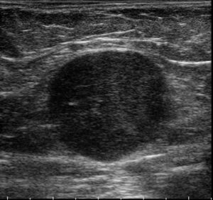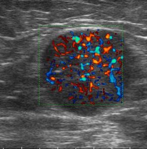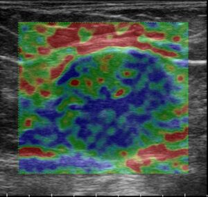
There is a real need for the creation and implementation of a simple tool for better communication between radiologists, clinicians and pathologists concerning lymph node (LN) assessment; particularly giving a clear signal and describing risk of cancer involvement.The LN-RADS scoring system, universal for any diagnostic modality (e.g., US, CT, MRI), realizes the idea of simplifying it lymph node classification.